Low-Grade Glioma
Low-grade gliomas are primary brain tumors that often occur in young and healthy adults following an initial seizure. These tumors require specialized surgery and advanced molecular testing to determine the most effective course of treatment. With modern medical therapies, many patients maintain a high quality of life for 10 years or more.
What Is Low-Grade Glioma (IDH-mutant Astrocytoma)?
A low-grade glioma is a slow-growing tumor that arises from glial cells — the support cells of the brain. Under the 2021 WHO classification, the diffuse low-grade gliomas most adults hear about are IDH-mutant astrocytomas (WHO grade 2 or 3) and IDH-mutant, 1p/19q-codeleted oligodendrogliomas (grade 2 or 3). They are grouped together because they share a founding mutation in the IDH1 or IDH2 gene.
These tumors disproportionately affect people in their 20s, 30s and 40s. Because they grow slowly and infiltrate normal brain without destroying it, the first sign is often a seizure in someone who feels otherwise healthy. 'Low-grade' does not mean harmless — over time, many transform into higher-grade cancers.
At a Glance
- Low-grade gliomas are slow-growing brain tumors that often present with a first-time seizure in a young adult
- The modern diagnosis is molecular: IDH1/IDH2 mutation and 1p/19q codeletion status define the tumor type and behavior
- How much tumor the surgeon safely removes is the single biggest modifiable factor in how long you live
- Awake craniotomy with brain mapping lets surgeons remove tumor from speech, language and motor areas without causing permanent deficits
- Vorasidenib, an IDH inhibitor pill, was FDA-approved in 2024 and can delay the need for radiation and chemotherapy in many patients
Most common presentations
- A first-time seizure — a full convulsion, brief staring spell, sudden unfamiliar smell or taste. Up to 80% of people with a low-grade glioma have a seizure at some point.
- Headaches that are new, persistent or worse in the morning.
- Subtle cognitive changes — trouble finding words, slower thinking, difficulty multitasking.
- Focal weakness, numbness, or clumsiness on one side.
- Language problems if the tumor is in or near the left hemisphere speech areas.
If you had a first-time seizure as an adult, you should have a brain MRI.
- Step 1: MRI with contrast. Classic low-grade gliomas appear as a bright area (on specific MRI settings like T2/FLAIR) that does not light up with contrast dye, gently expanding the normal brain tissue. We use advanced imaging to distinguish the tumor from a stroke, infection, or higher-grade tumor.
- Step 2: Surgical tissue diagnosis. Imaging alone cannot confirm the exact type of tumor. Our preferred approach is to perform a maximal safe removal of the tumor during the same operation that provides the tissue for diagnosis. If a larger surgery poses too much risk to your brain function, we can safely perform a minimally invasive needle biopsy.
- Step 3: Molecular classification. We thoroughly test every low-grade glioma for specific genetic markers: IDH1/IDH2 mutations, 1p/19q codeletion, ATRX/TP53/CDKN2A/B/TERT promoter status, and MGMT promoter methylation. For example, a CDKN2A/B deletion changes the classification of an IDH-mutant astrocytoma to a grade 4, guiding us toward a more specific treatment plan.
Astrocytoma, IDH-mutant (WHO grade 2 or 3)
IDH mutation but no 1p/19q codeletion; typically carries ATRX loss and TP53 mutation. Most common low-grade glioma in young adults. Median survival >10 years.
Oligodendroglioma, IDH-mutant and 1p/19q-codeleted (WHO grade 2 or 3)
The most favorable diffuse glioma. Median survival approaches 15-20 years. Especially responsive to radiation + PCV chemotherapy.
Glioblastoma, IDH-wildtype (WHO grade 4)
Included for contrast only. If older patient, enhancing mass, IDH-wildtype — this is NOT a low-grade glioma. Completely different treatment pathway.
Maximal safe resection is the foundation
More is better. In the landmark UCSF series, patients with ≥90% resection had 10-year overall survival of 91%, compared with 60% for patients with <90% resection.
Modern tools used: functional MRI and DTI, intraoperative neuronavigation, intraoperative MRI or ultrasound, 5-ALA fluorescence, and direct cortical/subcortical electrical stimulation.
Awake craniotomy with brain mapping
When tumor is in or near areas that control speech, language,or movement. Patient wakes during the critical part; a speech pathologist runs tasks while the surgeon maps. Fewer than 2% of patients have a permanent language deficit at 6 months.
Vorasidenib: a new option before radiation
Brain-penetrant inhibitor of mutant IDH1 and IDH2. In the INDIGO phase 3 trial, vorasidenib more than doubled progression-free survival (~27 months vs ~11 months with placebo). FDA-approved August 2024.
Radiation and PCV chemotherapy
For high-risk features: radiation followed by PCV chemotherapy (procarbazine, CCNU, vincristine). Based on RTOG 9802 — median OS was 13.3 years in the combined-therapy arm (vs 7.8 years for radiation alone), with benefit concentrated in IDH-mutant patients.
- Louis DN, et al. The 2021 WHO Classification of Tumors of the Central Nervous System. Neuro-Oncology. 2021;23(8):1231-1251. PMID: 34185076
- Mellinghoff IK, et al. Vorasidenib in IDH1- or IDH2-mutant low-grade glioma (INDIGO trial). NEJM. 2023;389(7):589-601. PMID: 37272516
- Buckner JC, et al. Radiation plus procarbazine, CCNU, and vincristine in low-grade glioma (RTOG 9802). NEJM. 2016;374(14):1344-1355. PMID: 27050206
- Smith JS, et al. Role of extent of resection in the long-term outcome of low-grade hemispheric gliomas. J Clin Oncol. 2008;26(8):1338-1345. PMID: 18323558
- Jakola AS, et al. Comparison of a strategy favoring early surgical resection vs watchful waiting in low-grade gliomas. JAMA. 2012;308(18):1881-1888. PMID: 23099483
- Sanai N, et al. Functional outcome after language mapping for glioma resection. NEJM. 2008;358(1):18-27. PMID: 18172171
- Louis DN, et al. The 2021 WHO Classification of Tumors of the Central Nervous System. Neuro-Oncology. 2021;23(8):1231-1251. PMID: 34185076
- Mellinghoff IK, et al. Vorasidenib in IDH1- or IDH2-mutant low-grade glioma (INDIGO trial). NEJM. 2023;389(7):589-601. PMID: 3
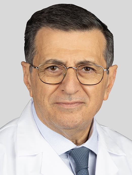
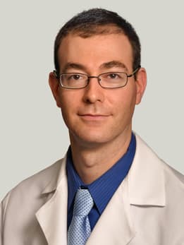
Our Specialists
Request an Appointment
We are currently experiencing a high volume of inquiries, leading to delayed response times. For faster assistance, please call 1-773-702-2123 to schedule your appointment.
If you have symptoms of an urgent nature, please call your doctor or go to the emergency room immediately.
* Indicates required field