Team of experts helps newborn survive rare fetal neck mass

A team of more than two dozen University of Chicago Medicine physicians and other clinicians saved a newborn — at birth — from a giant neck mass, illustrating the academic medical center's expertise in complex pediatric surgery and complicated obstetrical deliveries.
The drama, which unfolded in 2013, began when Sofia Espinoza arrived at her obstetrician’s office in Bloomingdale, IL, for a routine 24-week ultrasound on her second child. A few minutes into the appointment, her doctor noticed what appeared to be an abnormal growth on the baby’s neck.
“Usually when you go for an ultrasound, everyone’s so upbeat and eager to share the gender of the baby,” said Espinoza, of Addison, IL. “This time there was a scary silence in the room. I felt immediately that something must be wrong. My heart sunk when I was told the doctor wanted to speak with me in her office.”
A few days later, a more detailed ultrasound confirmed the mass on the baby’s neck was a cervical teratoma, a rare tumor that affects as few as 1 in 40,000 pregnancies. Although this one was benign, teratomas can hold serious implications for both mother and baby. Doctors informed Espinoza that there was a strong possibility that the tumor would compress the baby’s airway, which meant surviving birth would be an uphill battle.
Frightened and overwhelmed, Espinoza took to the web in search of more information, but found little. She was referred from obstetrician to obstetrician, each with few answers. Only one thing seemed clear: As the baby grew, so did the teratoma. At 28 weeks, the tumor measured 11 centimeters in diameter — nearly the size of the baby’s head.
“It was an incredibly stressful time,” Espinoza said. “I was moving from doctor to doctor. They all knew what it was, but they didn’t know what to do next. No one had experienced this. Everyone tried to remain hopeful, but I could see the nervousness in their faces. I had to start coping with the possibility that my baby may not make it.”
Espinoza was told the baby’s best chance for survival would be a specialized cesarean section delivery called an ex utero intrapartum treatment, or EXIT, procedure. During the procedure, a baby is partially delivered to expose the head and neck while continuing to receive oxygen from the mother via the placenta. A high level of skill is required to keep the uterus still and the placenta and umbilical cord intact while a surgeon works to insert a tube into the throat to clear an airway, allowing the baby to breathe once fully delivered.
Espinoza found the help she needed from physicians at UChicago Medicine in Hyde Park.
With the pending birth only a few weeks away, a multidisciplinary team of experts in obstetrics, pediatric surgery, pediatric otolaryngology, adult anesthesiology, pediatric anesthesiology, neonatology, radiology and nursing assembled to orchestrate the complicated delivery. They convened to review images, discuss potential scenarios and draw up a blueprint for the EXIT procedure that included timing, equipment and staffing needs, even the most ideal location for the delivery.
The birth was weeks in planning and required seamless coordination. Knowing the great risks involved with this procedure, the physician team wanted to anticipate any potential problem. To prepare, the entire team gathered in the OR for a simulation to make certain that everything was in place and every team member was prepared.
During a regular visit with maternal-fetal medicine specialist Deborah Boyle, MD, the week before the EXIT procedure was scheduled, a monitor revealed Espinoza was having contractions. Feeling no pain, she dismissed any concerns and headed home. At the strong urging of Boyle, Espinoza reluctantly returned to the hospital that same evening for close monitoring.
We’d been preparing for every possible scenario since the moment we learned about this case. There was no doubt we were ready. — Fuad Baroody, MD
This turned out to be a wise decision — just a couple of hours after Espinoza was settled in a bed, her water broke. Her son, John Carlos Guzman, was ready to make a Valentine’s Day debut.
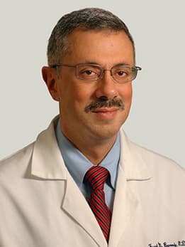
Fuad Baroody, MD, director of pediatric otolaryngology, received the page at roughly 3 a.m. In his decade at the University of Chicago Medicine Comer Children’s Hospital, the renowned head and neck surgeon had performed the critical task of securing an airway during several EXIT procedures and other cases involving complicated airway restriction. He knew no two cases were ever the same.
“I knew this would be one of our more complex cases,” said Fuad Baroody, MD, director of pediatric otolaryngology. “We’d been preparing for every possible scenario since the moment we learned about this case. There was no doubt we were ready.”
When Espinoza was wheeled into the operating room before dawn, more than 30 physicians and staff were waiting — one crew to tend to the mother and another devoted to the baby. Kenneth Nunes, MD, medical director of labor and delivery, joined Boyle for a final assessment of the uterus via ultrasound to determine the ideal location for the incision. They skillfully made an opening directly over the baby’s crown and cautiously withdrew his head to reveal the large mass dominating the right side of his neck and face.
Baroody then went to work executing the intubation and found that the size and position of the tumor presented a challenge: The windpipe was displaced far to the left. To overcome this obstacle, he elected to use the tiniest of light tubes, just 2 millimeters in diameter, to guide a path to the trachea. A test confirmed the breathing tube had been successfully inserted. Baroody gave the green light for the delivery to be completed.
John Carlos’ first hurdle now was behind him. Neonatologists, led by executive vice chairman of pediatrics Michael Schreiber, MD, swiftly moved the baby to an adjacent room to check his vitals, place intravenous lines and ensure he was stable enough to make the short trip on the medical campus from Bernard A. Mitchell Hospital to neighboring Comer Children’s.
Just hours later, John Carlos underwent a delicate surgery to remove the tumor. For Baroody and the Comer Children's pediatric surgery team, a specialist in congenital anomalies, the biggest challenge was to cut away the large teratoma, now 12 centimeters in diameter, without damaging healthy tissue and the hair-like nerves nearby that would allow the baby to swallow, cry or smile.
In an operation that lasted more than five hours, the tumor was successfully removed. John Carlos proved strong, surviving the surgery and continuing to grow and defy any concerns during his three-week stay in the neonatal intensive care unit. Aside from a scar on the neck and the extra skin once filled by the tumor, there are no traces of the serious medical challenges he once faced. Doctors say he's expected to live a normal, healthy life.
“His recovery has been truly amazing,” Espinoza said. “When I saw the pictures of him with the tumor, I gasped. You just can't prepare yourself for that. I know we were lucky to find the right doctors.
“When I was searching everywhere for positive stories about teratomas this large, there were none," the mother added. "That’s why I want to share my experience with other parents.”