
Gut-pancreas immune crosstalk may offer new therapeutic strategies for pancreatic cancer
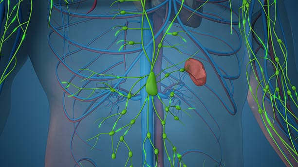
Lymphatic drainage in the abdomen
In a recent study published in Cell Reports, researchers at the University of Chicago reported that immune responses produced in lymph nodes shared by the liver, pancreas and intestine shape T-cell tolerance.
They found that pathological events such as intestinal viral infections can reprogram pancreas or liver-reactive T cells into inflammatory cells within those shared lymph nodes. This cross-organ immune rewiring also increased T-cell infiltration into pancreatic tumors and improved tumor control, pointing to potential new approaches for hard-to-treat pancreatic cancers.
The immune system is often described as a bodywide defense network, but it does not function uniformly across tissues. Immune cells that enter organs such as the lung, gut, liver, pancreas or brain adapt to local cues and develop tissue-specific immunity. Nearby lymph nodes act as regional command centers, helping determine whether to mount an inflammatory response or promote tolerance.
A key question is whether immune responses generated in one organ can shape responses in others through shared lymphatic drainage.
“We usually think of lymph nodes as guards for specific sites,” said Daria Esterhazy, PhD, senior author of the study and Assistant Professor of Pathology at the University of Chicago. “But some lymph nodes are shared between different organs and if something is going on in the gut, and that lymph node is also draining the liver and pancreas, it can affect how the immune system reacts to those other organs.”
Tissue-specific immune tolerance
Immune tolerance is a process that prevents T cells from attacking the body’s own tissues. T cells are trained to tolerate self-antigens in a unique and tissue-specific way so that they ignore harmless or self-derived signals and mount a strong response against infection or cancer.
Using mouse models, Esterhazy’s team compared how the immune system responded to the same model antigen when it came from different tissues such as the liver, pancreas or intestine and when it appeared in different forms. They found that the fate of T cells depended not just on the antigen itself, but on where it came from, how much of it was present and how accessible it was to immune cells.
In the liver and pancreas, antigens hidden inside cells were often ignored by the immune system, a process known as immunological ignorance. When large amounts of secreted antigen came from the liver, the immune system eliminated the reactive T cells through clonal deletion. In contrast, in the pancreas and intestine, secreted antigens pushed T cells toward becoming regulatory T cells, or Treg cells, which help suppress immune responses and prevent autoimmune damage.
“The same self-antigen can produce very different T-cell fates depending on whether it comes from the liver, pancreas or intestine,” Esterhazy said. “T-cell tolerance depends on context like the source, location and amount of antigen, and the condition of the tissue.”
Immune crosstalk between organs
The team conducted experiments to understand what happens when one organ in the shared network becomes inflamed. They induced an intestinal viral infection using reovirus, which triggers a Type 1 antiviral immune response.
Even though the infection was in the gut, it changed the behavior of T cells that recognized pancreatic or liver antigens. In shared lymph nodes, those T cells became more inflammatory and cytotoxic, making them more capable of killing cells. They then migrated into the pancreas or liver and caused autoimmune-like tissue damage.
Interestingly, this effect was specific to lymph nodes that were shared by the inflamed tissue and the antigen-producing tissue, emphasizing that the immune crosstalk depended on anatomical connection through lymphatic drainage.
This matters because the pancreas shares lymphatic drainage with parts of the gut. The upper intestine is constantly exposed to food, microbes and environmental signals, and its immune environment is often geared toward tolerance. That helps prevent harmful responses to food, but it may also make it harder for the immune system to mount strong antitumor responses against pancreatic cancer.
“With respect to pancreatic cancer, sharing lymph nodes with the gut is overall not ideal,” Esterhazy said. “The intestinal lymph node is very tolerance-promoting, which is great news for preventing food allergy and autoimmunity, but it may be fatal for cancer.”
Reprogramming the lymph node, not the pancreas
Pancreatic cancer is notoriously resistant to many immunotherapies. One reason may be that tumor antigens draining from the pancreas enter lymph nodes that are biased toward tolerance rather than immune attack.
The researchers tested whether gut-driven inflammation could change that environment. The idea was not to inflame the pancreas directly, which can be dangerous, but to use the gut-pancreas lymph node connection to alter how T cells are instructed to fight pancreatic cancer.
“The pro-inflammatory signal comes from the gut, and the antigen comes from the pancreas,” Esterhazy said. “They meet in that shared lymph node, and that allows you to reprogram the antitumor response; essentially, we are reprogramming the lymph node without directly perturbing the pancreas.”
In one pancreatic cancer model, where the target antigen was broadly expressed in pancreatic tissue, intestinal reovirus made disease worse. The activated T cells attacked not only tumor-related cells but also antigen-expressing pancreatic tissue, leading to damage and accelerated tumor progression. This effect was driven largely by cytotoxic T cells, the immune cells best known for killing infected or abnormal cells.
But in another model, where the antigen was restricted to tumor cells, gut infection improved tumor control. Intestinal reovirus increased T-cell infiltration into pancreatic tumors and reduced tumor burden. This occurred only when the tumor antigen was secreted and accessible to immune cells in the shared lymph nodes. Antigens hidden inside tumor cells did not produce the same benefit.
“There is a problem if the antigen from the pancreas is not specific to the tumor,” Esterhazy said. “Then you are going to destroy not only the tumor cells, but also normal cells, which can lead to pancreatitis.”
A possible path toward safer immunotherapy
The findings highlight that it may be possible to deliver signals through the gut that make shared lymph nodes more supportive of antitumor immunity. The study further suggests that the safest and most effective strategies would need to focus on tumor-specific antigens, targets present on cancer cells but not normal pancreatic tissue and on antigens that immune cells can readily sample.
The work also adds to a growing understanding that the immune system’s geography matters.
“Delivering something through the gut route that renders the lymph node more pro-inflammatory might be beneficial,” Esterhazy said.
Lymph nodes are not interchangeable, and organs that share lymphatic drainage may influence one another in unexpected ways. For pancreatic cancer, that could be especially important. Rather than targeting the tumor alone, future therapies may also need to consider the immune environment where antitumor responses begin.
“This study shows that shared lymph nodes can maintain tolerance, trigger tissue damage or support antitumor immunity depending on the context,” Esterhazy said. “Understanding that context may help us design better ways to guide immune responses in pancreatic cancer.”
The study, “Tissue-specific tolerance mechanisms and lymph node co-drainage shape T cell immunity in the upper digestive system and pancreatic cancer progression,” was supported by funds from the Pancreatic Cancer Action Network, the Cancer Research Foundation, the National Institutes of Health, and startup funds from the University of Chicago.
Additional authors include Yixuan Zhou, Peter Wang, Emily Schaffer, Macy Komnick, Hailey Brown, Colin Sheehan, and Alexander Muir from the University of Chicago; Gwen Taylor, Kay Fiske, and Terence S. Dermody from the University of Pittsburgh School of Medicine.
UChicago Medicine and the Biological Sciences Division continue to be at the forefront of cancer care and research. In April 2027, UChicago Medicine will open the AbbVie Foundation Cancer Pavilion, the state’s first freestanding cancer pavilion, to bring advanced diagnostics, innovative treatments, translational discoveries and comprehensive support to patients and the community.

UChicago Medicine Comprehensive Cancer Center
UChicago Medicine is designated as a Comprehensive Cancer Center by the National Cancer Institute, the most prestigious recognition possible for a cancer institution. We have more than 200 physicians and scientists dedicated to defeating cancer.
Learn More About the Comprehensive Cancer Center