Nuclear medicine's potential to combat cancer
Nuclear medicine has been a powerful approach in cancer diagnosis and treatment for over 50 years. Just recently researchers and physicians have pushed the field forward with the development of new tools for precision medicine.
One might say that the field of modern nuclear medicine was born at the University of Chicago. In fact, the world’s first nuclear reactor was used to create the first controlled, self-sustaining nuclear reaction at University of Chicago in 1942, launching the nuclear era.
As a research institution and a hospital, there was always an immense interest in using this technology for the diagnosis and treatment of cancer. This led to the opening of the Argonne Cancer Research Hospital in 1954 on the UChicago campus. Argonne Cancer Research Hospital was the largest facility ever built for the purpose of cancer research and treatment using nuclear medicine.
An early team of UChicago researchers including Katherine Austin Lathrop, BS, MS, Paul Harper, MD, Robert Beck and Alex Gottschalk, MD, launched the field of modern nuclear medicine with the development of an imaging technique using a radiotracer, a radioactive molecule that binds to cancer in the body, which is the most commonly used medical radioisotope today, almost 60 years later.
This imaging technology could help clinical teams determine whether a cancer is aggressive and inform treatment decisions.
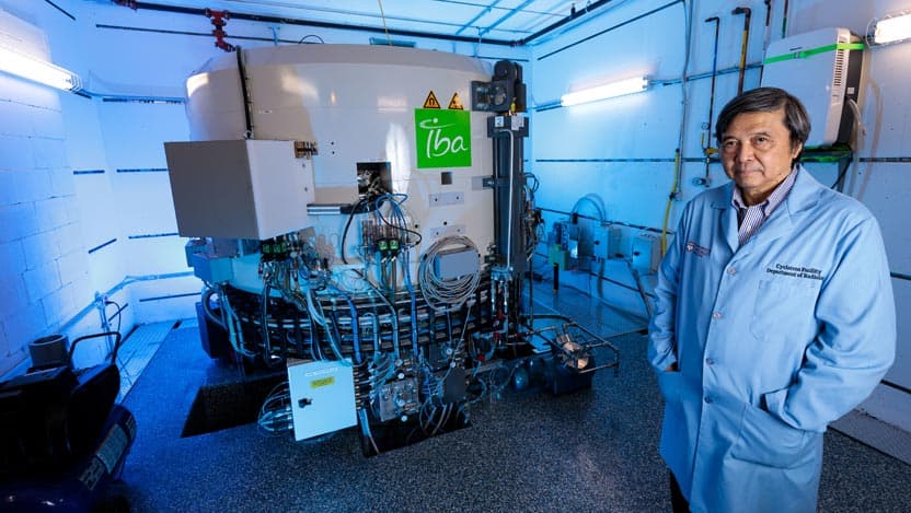
For progress to continue, the University installed a giant machine called a cyclotron in 1968. The cyclotron is used to create radioactive particles, or isotopes, which are used to produce radiopharmaceuticals. Having direct access to a cyclotron allowed for steady progress in the field of nuclear medicine for decades.
In recent years, triple-negative breast cancer and in other cancers. Moellering, Chen and their teams demonstrated that this new type of radiotracer can find and label aggressive cancer cells in whole-body imaging in an animal model.
“This imaging technology could help clinical teams determine whether a cancer is aggressive and inform treatment decisions,” Moellering said. “This enzyme is elevated in many different types of aggressive cancer, so if it works to track aggressive breast cancer, it may have utility in tracking many other types of aggressive cancer.”
Extraordinary achievements in the use of nuclear medicine to diagnose and treat cancer is a part of the history of UChicago. With the recently installed modern cyclotron, a new era of discovery and research has been initiated for UChicago Medicine Comprehensive Cancer Center clinicians and investigators. Novel radiotracers have the potential to evolve the field toward precision medicine, improving results and quality-of-life for cancer patients.