University of Chicago scientists advance heart tissue research as finalists in NASA competition
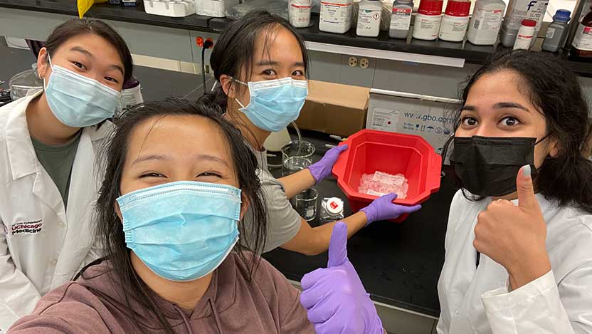
Undergraduate researchers show off some of their heart tissue samples in the lab.
When it came to the NASA Vascular Tissue Challenge, the bar was high: grow a piece of vascularized tissue that is over 1 centimeter thick, and make sure that at least 85% of the cells within the tissue survived over 30 days.
That might not sound like much, but to put that in perspective, “Consider that the best we’ve been able to do so far was just a couple of millimeters,” said Narutoshi Hibino, MD, PhD, Associate Professor of Surgery at the University of Chicago Medicine. “So, NASA knew that this is a very challenging thing to do.”
The challenge: growing an organ…in space
The Vascular Tissue Challenge offered a $500,000 prize to be split among the first three teams to “successfully create thick, metabolically-functional human vascularized organ tissue in a controlled laboratory environment.” Teams also had to submit proposals on how to further advance their research through an experiment that could be conducted in microgravity on the International Space Station.
An important component of the project is the goal of developing new, self-sustaining technology that can be used by astronauts to conduct studies on the effects of space travel and low gravity on the human body. Understanding these key details will be important for determining the feasibility and safety of long-term space travel and perhaps someday colonizing other planets.
Despite the tall order, Hibino and his team were undeterred, even as the COVID-19 pandemic led to major changes in the laboratory, causing research backups and priority changes. Out of more than 20 institutions that originally signed up to compete in the competition, the Hibino lab was one of just five teams remaining by the time the final experiments launched in the summer of 2021.
The challenge was not completely out of the realm of possibility for the team — the Hibino lab works on engineering cardiac tissue using 3D and bioprinting technology. But simply creating the tissue is not the only hurdle that has to be overcome.
“The challenge is in the vascularization,” said Hibino. “When tissue is thicker than a fraction of a millimeter, the cells on the inside of the tissue become deficient in oxygen and they die. To create thick tissues, we need to incorporate a vascular tree to support the tissue. But that’s been very challenging, and we don’t have the technology yet.”
Vascularization — the presence of blood vessels — provides living tissue with essential nutrients and oxygen to keep it thriving. In the Hibino lab, that translates to building 3D tissues using bioprinters to produce round clumps of tissue called spheroids. The research team, which consisted primarily of undergraduate students, made several trial runs ahead of the final challenge.
“There were good surprises and bad surprises,” said Hibino. “We have changed the system many times, trying different ways to see what works best, and we’ve made some interesting discoveries that way. But we’ve also dealt with contamination issues, and our work was complicated when undergraduates could not come to campus because of the COVID-19 pandemic.”
The final test
Finally, on May 12, 2021 the final project was launched.
The team designed a system that incorporated two different kinds of human cells: induced pluripotent stem cells (iPSCs) and cardiac fibroblasts. The iPSCs are stem cells that can develop into different cell types based on which chemical signals they are exposed to during growth. They coaxed the cells into spheroids before transferring the resulting cell clumps into a 3D printed mold to further expand. The mold was placed in a custom chamber designed to allow flow of the growth media supporting the cells, providing them with continuous nutrients and oxygen. From there, they pushed the iPSCs to develop into cardiomyocytes, another type of heart cell, using a special cocktail of molecules added to the growth media. Finally, they encouraged vascularization by seeding the tissue with endothelial cells collected from the human umbilical vein.
The cells were then allowed to grow undisturbed for 30 days in their custom chamber.
While the researchers were able to keep their tissue samples alive for the full 30 days, they were not able to hit every metric for the challenge.
“Our tissue size was small,” said Hibino. “We only achieved about 80% of the required 1 cubic centimeter. And, the most difficult part — we were not able to achieve functionality of our tissue, meaning that our cardiac tissue did not beat. This would have been very difficult to do. It’s a little easier to do with some other tissues, such as liver, but we decided not to compromise.”
New directions for the future
Ultimately, while the work did not garner a portion of the prize money, the researchers are proud of what they accomplished, and of what this kind of research means for the future of their field.
“To create human tissue, or any kind of organs, the biggest challenge is getting that 3D structure,” said Hibino. “We can’t yet create a heart or a liver, which is why transplantation is our only current solution. So, it’s not just about overcoming the challenge of developing this specific technology, but it’s also about changing the future of science and medicine.”
In addition, the challenge has inspired new research projects in the lab. “Now that we’re through this game, we’ve got about 20 undergraduate students in the lab, and each of them has a unique project to work on because of all the questions that came up during the challenge that need to be answered,” said Hibino. “This has given us a huge opportunity to advance all of our research work, like a jumping off project. Sometimes it can be difficult to make a big research plan, but with this project we were able to advance a lot of questions.”
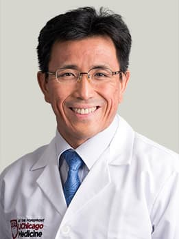
Narutoshi Hibino, MD, PhD
Narutoshi Hibino, MD, PhD, is a highly respected surgeon with extensive experience in pediatric cardiac surgery and performs a wide range of complex procedures for patients with congenital heart disease.
View Dr. Hibino's physician profile