Beyond joint replacement: Restoring cartilage to its original condition

Joint replacements are a great option for patients with debilitating injuries or arthritis. More than one million Americans have a hip, knee or shoulder replaced each year, and the procedures have become so routine that many patients are up on their feet and walking a few days after surgery.
But they're not perfect. It's still invasive surgery, and patients run the risk of infections and other complications. Plus, the new joint can wear down after many years, requiring additional repair procedures.
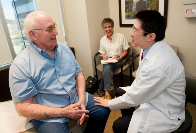
Lewis Shi, MD, an orthopedic surgeon at the University of Chicago Medicine who specializes in shoulder and elbow injuries, says that even though joint replacements have been perfected over the last few decades, surgeons should still view them as a last resort. "Joint replacement is getting better and better, but it's not getting better at a rate where you can overcome an aging population that continues to be very active," he said.
Shi's research focuses on the mechanics and cell-level basis of shoulder injuries and arthritis, including properties of cartilage that affect healing. Joint surfaces are covered with a smooth type of cartilage, called hyaline cartilage, which enables them to move freely. If that cartilage is damaged though, it's gone for good.
Surgeons have several surgical techniques at their disposal to repair damage and encourage cartilage regeneration. Microfracture surgery, for example, can be used when a patient has a small, focused area of damage where cartilage has been worn away to the bone. Surgeons drill small holes into the bone to encourage bone marrow to come to the surface and regrow cartilage. But the cartilage that regrows isn't the same smooth hyaline cartilage-it's thick fibrocartilage, like scar tissue on the skin, which can interfere with movement in the joint and cause further damage.
Another option is called the OATS procedure, or osteochondral autograft transfer system. Surgeons take a round, plug-shaped piece of bone and cartilage from a healthy part of the joint and transplant it to the damaged area, like doing a skin graft to cover a patch of burned skin.
A third, more advanced procedure called autologous chondrocyte implantation (ACI) involves harvesting healthy cartilage and culturing the cells that produce new cartilage, called chondrocytes. Surgeons transplant the new cells back into the damaged area in a gel or stitch them into place under a membrane to encourage regrowth.
The behavior of chondrocyte cells in cartilage is still rather mysterious though, partly because they don't live very long in experimental settings. Researchers can culture them, but they don't last long enough to see the results of experiments.
Shi and Tong-Chuan He, PhD, an associate professor of surgery, are doing research on how to prolong the life of chondrocytes in the lab. If scientists could get these cells to live indefinitely, they could try things like growing them on three-dimensional scaffolds that could reproduce cartilage tissue, or see how they respond to different forces of gravity in a weight-bearing joint. They could do genetic analysis, or treat them with different growth factors to see how the chondrocytes change or grow over time.
Shi says better understanding of how chondrocytes work could improve existing techniques like microfracture or ACI to encourage the growth of smooth hyaline cartilage. It might also provide ways to spot damage to cartilage and development of arthritis early enough to intervene before joint replacement becomes the only option.
"Even though joint replacement is a good solution, at a population level we cannot abandon other techniques," Shi said. "We still want to put patients off replacements as long as we can."

Lewis Shi, MD
Lewis Shi, MD, is an orthopaedic surgeon who specializes in shoulder and elbow injuries.
Learn more about Dr. Shi
